
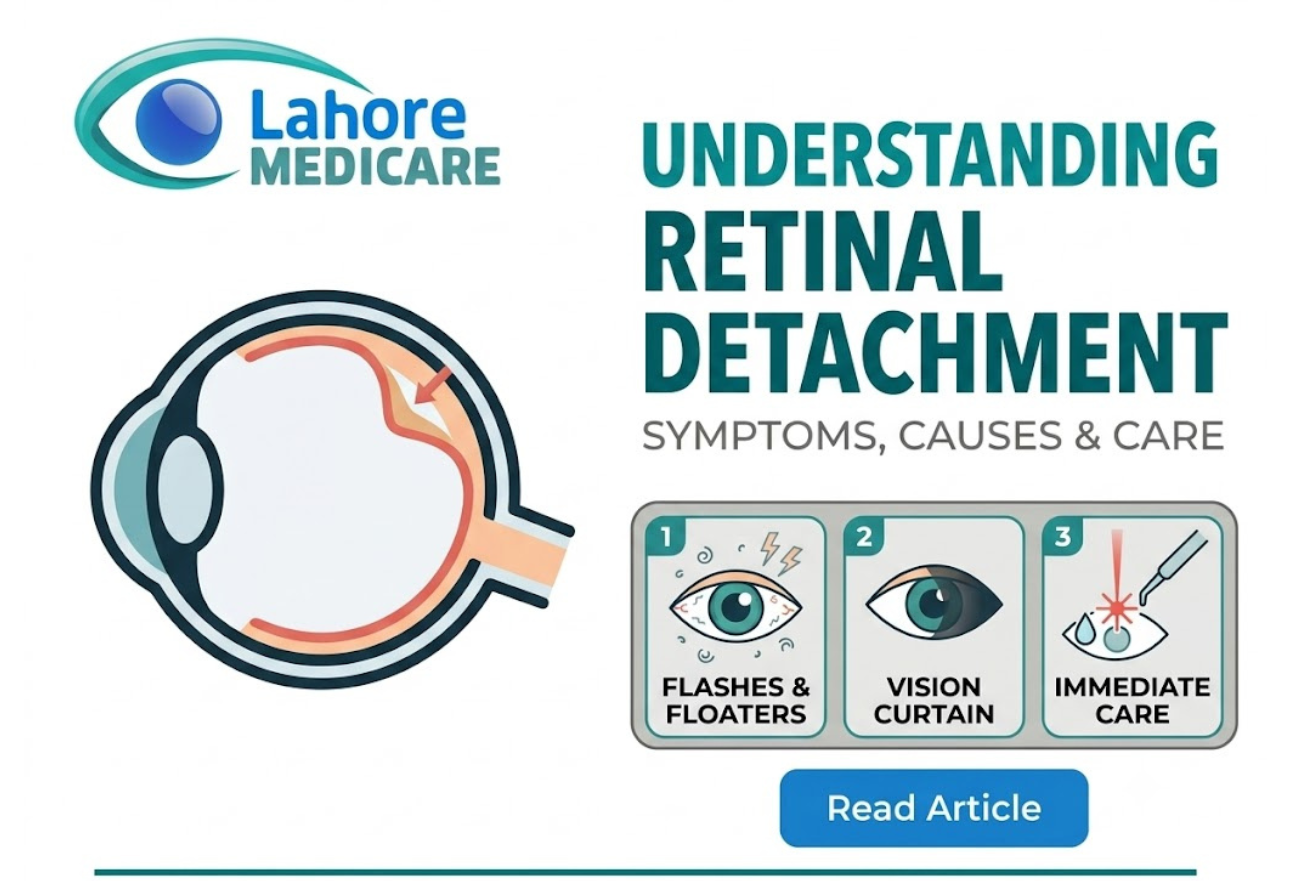
Vision relies on a smooth, uninterrupted transmission of light signals from the eye to the brain. At the center of this process is the retina, a thin, light-sensitive layer of tissue lining the inside back wall of the eyeball. The retina contains millions of photoreceptor cells (rods and cones) that receive visual focus, organize the information, and send it to the brain through the optic nerve.
When the retina pulls away or separates from its underlying supportive tissue and blood vessels, it creates a medical emergency known as retinal detachment. Without an active blood supply, retinal cells quickly starve of oxygen. Left untreated, a detached retina can spread across the entire posterior chamber, leading to permanent, severe vision loss or complete blindness.
The Three Primary Causes of Retinal Detachment
Retinal detachment can occur due to aging, structural changes, trauma, or secondary ocular diseases. Ophthalmologists classify the condition into three distinct mechanisms:
1. Rhegmatogenous Retinal Detachment (Most Common)
This type occurs when a small tear or break develops in the retina. The clear, gel-like substance that fills the center of the eye (the vitreous humor) leaks through the tear and pools underneath the retina. As fluid accumulates, it gradually lifts the retina off the underlying retinal pigment epithelium (RPE). Age-related vitreous shrinkage is the leading cause of these tears.
2. Tractional Retinal Detachment
Tractional detachment happens when abnormal scar tissue grows on the retina’s surface, pulling it away from the back of the eye. This form is most commonly seen in patients with advanced, unmanaged diabetic retinopathy, where fragile, abnormal blood vessels leak and scar over time.
3. Exudative (Serous) Retinal Detachment
In exudative detachment, fluid accumulates beneath the retina without any tears or breaks present. This fluid buildup is usually triggered by inflammatory diseases, vascular malformations, eye trauma, or intraocular tumors.
Recognizing Early Warning Symptoms
Retinal detachment itself is entirely painless because the retina lacks pain fibers. However, it almost always produces distinct visual warning signals before total detachment occurs. You may need to test each eye individually to notice subtle early changes.
Key warning signs include:
- Sudden Increase in Floaters: The sudden appearance of numerous tiny specks, threads, or cobweb-like shapes drifting across your field of vision.
- Flashes of Light (Photopsia): Brief, bright flashes or streaks of light, especially visible in dark environments or when moving your eyes.
- A Dark “Curtain” or Shadow: A gradual dark shadow or veil falling across your peripheral (side) vision that expands toward the center.
- Blurred or Distorted Vision: Straight lines appearing wavy, curved, or out of focus as the central macular area becomes affected.
Crucial Warning: If you suddenly experience flashes, a flood of new floaters, or a shadow over your field of vision, seek immediate emergency eye care. Prompt medical intervention is essential to preserve your sight.
Risk Factors to Consider
While retinal detachment can happen to anyone, certain factors significantly increase your susceptibility:
- Extreme Myopia (Nearsightedness): Severe nearsightedness stretches the physical eyeball, making the peripheral retina thinner and more prone to tears.
- Previous Eye Surgery: Prior complex procedures, such as high-risk cataract removal, can alter vitreous stability.
- Severe Ocular Trauma: Direct impacts or blunt injuries from sports, accidents, or physical trauma.
- Family or Personal History: Having a history of retinal tears in one eye increases the risk of developing detachment in the other.
- Underlying Retinal Diseases: Chronic conditions like advanced diabetic eye disease or inflammatory retinal disorders.
For individuals navigating complex ocular conditions, obtaining a diagnostic evaluation at a specialized facility like Lahore Medicare helps identify peripheral tears before full detachment occurs.
Immediate Clinical Treatments
Retinal tears and full detachments cannot be treated with medication or eye drops. Surgery is the only effective way to repair the retina and restore blood supply.
Emergency Procedures for Retinal Tears
If caught early when only a small retinal tear or hole exists without full detachment specialists use non-invasive laser or freezing procedures:
- Photocoagulation (Laser Surgery): An argon laser beam is directed through the pupil to create microscopic burns around the retinal tear. The resulting pinpoint scar tissue welds the retina back to its underlying layer.
- Cryopexy (Freezing): A freezing probe applied to the outer surface of the eye freezes the tissue around the tear, creating a scar that seals the retina in place.
Surgical Repairs for Full Retinal Detachment
When fluid has already separated the retina from the eye wall, advanced surgical intervention is required:
- Pneumatic Retinopexy: The surgeon injects a small gas bubble into the vitreous cavity. The bubble expands and presses against the detached area, pushing it back against the wall of the eye while laser or cryotherapy seals the tear.
- Scleral Buckling: A tiny silicone band or sponge is attached around the outside wall of the eye (sclera). This gently indents the eye wall to relieve the traction pulling on the retina.
- Vitrectomy: The surgeon removes the clear vitreous gel pulling on the retina, clears any internal scar tissue, and fills the cavity with air, gas, or silicone oil to hold the retina in its proper anatomical position.
Patients requiring advanced surgical intervention can access specialized care for complex retinal conditions and emergencies to preserve functional vision. Furthermore, utilizing state-of-the-art ophthalmic diagnostic and surgical procedures ensures high precision and better recovery outcomes.
Frequently Asked Questions
Is retinal detachment painful?
No. Retinal detachment is completely painless because the retina does not contain pain receptors. You will only notice visual symptoms like floaters, flashes, or a dark shadow in your vision.
Can a detached retina heal on its own?
No. A detached retina cannot repair itself. Without immediate medical or surgical intervention, the retina will continue to detach, leading to irreversible vision loss.
What is the difference between a retinal tear and a retinal detachment?
A retinal tear is a small hole or rip in the retinal tissue. A retinal detachment occurs when fluid seeps through that tear and lifts the retina away from the back wall of the eye.
How quickly do I need treatment for a detached retina?
Retinal detachment is a medical emergency. You should seek immediate treatment within 24 to 48 hours to prevent permanent damage, especially if the central area of vision (the macula) is still attached.
What causes flashes of light in your vision?
Flashes occur when the vitreous gel inside the eye pulls on or rubs against the sensitive retina. This physical traction stimulates the retina to send light signals to the brain.
Are people with high myopia at higher risk?
Yes. Nearsighted eyes are elongated, which stretches the retina and makes it thinner, weaker, and more susceptible to developing peripheral tears.
What is the recovery period after retinal detachment surgery?
Initial recovery takes 2 to 4 weeks, during which you may need to maintain a specific head position if a gas bubble was used. Full visual stabilization can take several months.
Can diabetic retinopathy cause retinal detachment?
Yes. Advanced diabetic retinopathy can cause abnormal blood vessels to grow and scar on the retina. This scar tissue contracts and creates traction, pulling the retina off the eye wall.
How do doctors diagnose a detached retina?
Ophthalmologists perform a dilated eye exam, using specialized indirect ophthalmoscopes and visual ultrasound (B-scan) to inspect the retina and locate tears or fluid buildup.
Can you prevent retinal detachment?
While you cannot always prevent it, wearing protective eyewear during sports, getting regular dilated eye exams, and seeking immediate care for new floaters or flashes drastically reduce your risk.