
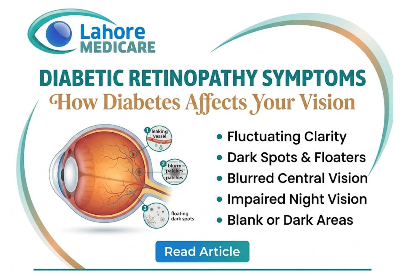
Diabetes is a complex systemic condition that impacts blood vessels throughout the human body. One of its most severe and prevalent long-term complications occurs deep inside the eye within the retina, the light-sensitive layer of neural tissue lining the back of the eyeball. When consistently high blood sugar levels damage the tiny microvascular capillaries supplying the retina, it leads to a condition known as diabetic retinopathy.
Because early-stage diabetic retinopathy often progresses silently without causing noticeable pain or immediate visual changes, many individuals remain unaware of the developing vascular damage until advanced complications threaten their sight.
How High Blood Sugar Damages Your Retina
The retina requires a continuous, stable supply of oxygenated blood to process visual signals and send them along the optic nerve to the brain. Prolonged high blood glucose levels disrupt this supply through two distinct structural mechanisms:
- Capillary Blockage and Microaneurysms: Over time, glucose spikes weaken the inner walls of delicate retinal capillaries. These weakened spots balloon outward into tiny pouches called microaneurysms, which eventually leak fluid, lipids, and blood into surrounding retinal tissue.
- Ischemia and Neovascularization: As damaged capillaries close off entirely, areas of the retina become starved of oxygen (ischemic). In response, the eye triggers the growth of new blood vessels (neovascularization). However, these newly formed vessels are exceptionally fragile and prone to rupturing, spilling blood directly into the clear vitreous gel of the eye.
Stages of Diabetic Retinopathy
Diabetic retinopathy generally progresses through two main clinical stages:
Non-Proliferative Diabetic Retinopathy (NPDR)
This is the early stage of the disease. The tiny retinal blood vessels swell, leak fluid, and develop microaneurysms. NPDR is categorized as mild, moderate, or severe based on the extent of vascular blockage and hemorrhages. During this stage, fluid can also collect in the macula the central focus area of the retina causing diabetic macular edema (DME), which leads to central blurriness.
Proliferative Diabetic Retinopathy (PDR)
This is the advanced, severe stage of the disease. Triggered by widespread retinal oxygen deprivation, fragile new blood vessels sprout across the retina and into the vitreous cavity. Because these vessels bleed easily, they can trigger massive vitreous hemorrhages, form contractile scar tissue, and cause tractional retinal detachment.
Recognizing Key Symptoms and Warning Signs
In the earliest phases of NPDR, you may not experience any obvious symptoms. As the disease advances, however, physical damage to the retinal microvasculature produces clear visual disruptions:
- Fluctuating Vision Clarity: Vision that shifts unpredictably between clear and blurry depending on daily blood sugar levels.
- Dark Spots or Floaters: Sudden streaks, dark spots, or cobweb-like shapes drifting across your vision caused by micro-bleeds into the vitreous humor.
- Blurry or Distorted Central Vision: Difficulty reading, recognizing faces, or seeing fine detail due to fluid accumulation in the macula.
- Impaired Night Vision and Color Perception: Difficulty navigating in low light or noticing that colors appear washed out and faded.
- Blank or Dark Areas: Patches of partial or total vision loss within your visual field.
Because visual symptoms often appear only after significant vascular damage has occurred, receiving a routine comprehensive screening at a tertiary care facility like Lahore Medicare provides crucial early detection before irreversible damage sets in.
Diagnosis and Modern Treatment Options
Ophthalmologists diagnose diabetic retinopathy using specialized diagnostic tools, including dilated fundus examinations, optical coherence tomography (OCT) to measure macular swelling, and fundus fluorescein angiography (FFA) to map leaking vessels.
While damaged retinal tissue cannot always be fully restored, modern treatments can halt disease progression and preserve functional vision:
- Anti-VEGF Injections: Medications injected directly into the eye block Vascular Endothelial Growth Factor (VEGF), reducing fluid leakage and shrinking abnormal, fragile blood vessels.
- Laser Photocoagulation: Focal laser treatment seals leaking retinal vessels, while pan-retinal photocoagulation (PRP) uses laser spots to shrink widespread abnormal vessels across the peripheral retina.
- Vitrectomy Surgery: An advanced surgical procedure used in proliferative stages to clear dense vitreous blood clots and peel away tractional scar tissue holding the retina.
For patients experiencing advanced diabetic vascular changes or macular swelling, receiving specialized care for diabetic eye diseases and conditions ensures long-term preservation of functional sight. Patients needing targeted surgical intervention or intravitreal therapies can benefit from advanced ophthalmic procedures designed to restore stability to fragile retinal blood vessels.
Frequently Asked Questions
Can diabetic retinopathy be reversed?
While structural damage to retinal nerves cannot be completely reversed, early-stage leakage and swelling can be significantly reduced with tight blood sugar control, anti-VEGF injections, and laser treatments to prevent further vision loss.
How often should diabetic patients get an eye exam?
Individuals with diabetes should undergo a comprehensive dilated eye exam at least once a year. If you have already been diagnosed with diabetic retinopathy, your specialist may recommend screenings every 3 to 6 months.
What is the difference between NPDR and PDR?
Non-proliferative diabetic retinopathy (NPDR) is the early stage where blood vessels leak fluid and swell. Proliferative diabetic retinopathy (PDR) is the advanced stage where fragile, abnormal new blood vessels grow on the retina and risk rupturing.
What is diabetic macular edema (DME)?
Diabetic macular edema occurs when leaking blood vessels cause fluid to accumulate in the macula the center of the retina responsible for sharp, straight-ahead vision leading to severe central blurring.
Can controlling blood sugar prevent diabetic retinopathy?
Yes. Maintaining strict control over your blood glucose levels, HbA1c, blood pressure, and cholesterol significantly reduces the risk of developing diabetic retinopathy and slows its progression.
Are anti-VEGF injections painful?
No. Before administering anti-VEGF injections, the eye is thoroughly numbed with anesthetic drops. Patients typically feel only mild pressure during the quick procedure.
What causes sudden floaters in diabetic patients?
Sudden floaters in diabetic patients are often caused by tiny blood leaks from fragile new vessels into the vitreous gel. A sudden flood of floaters requires urgent evaluation by a retina specialist.
Does diabetic retinopathy affect both eyes?
Yes. Because diabetes is a systemic condition that affects blood vessels throughout the body, diabetic retinopathy almost always develops in both eyes, though the severity may vary between eyes.
How does laser treatment help diabetic retinopathy?
Laser photocoagulation seals actively leaking blood vessels and reduces the retina’s demand for oxygen, which stops the growth of dangerous, fragile new vessels.
What are the early warning signs of diabetic eye damage?
In its earliest stages, there may be no warning signs at all. As it progresses, early signs include mild vision fluctuations, dark floating spots, blurred central vision, and difficulty reading in low light.