When discussing vision health, attention almost always centers on the visible parts of the eye, such as the cornea, lens, or retina. However, the delicate structures that allow the eye to focus, lubricate, and move reside within a protective biological housing called the orbital cavity. When disease, inflammation, or abnormal tissue growth develops within this housing, it disrupts both the mechanical function of the eye and the surrounding facial architecture.
Understanding orbital diseases begins with recognizing how closely packed these structures are. Because the eye socket is a rigid, bony space with limited room to expand, even minor swelling or tissue growth creates significant internal pressure, leading to distinct visual and physical symptoms.
What Is the Orbital Cavity?
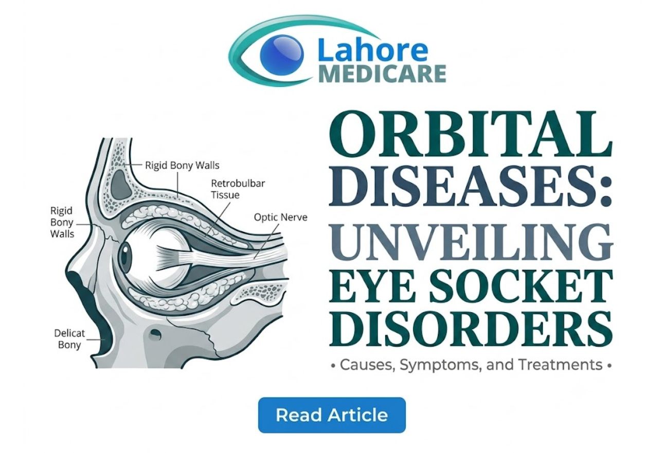
The orbital cavity (eye socket) is the bony cavity that encloses the bulb and accessory organs of the eye, including the ocular muscles, lacrimal glands, nerves, vessels, and retrobulbar adipose tissue. Each component plays a vital role in maintaining eye health:
- Ocular Muscles: Six specialized muscles that coordinate precise, multi-directional eye movements.
- Lacrimal Glands: Secretory glands located in the upper outer portion of the orbit that produce tears to lubricate the ocular surface.
- Nerves and Blood Vessels: Intricate networks, including the optic nerve, that transmit sensory data to the brain and supply oxygenated blood.
- Retrobulbar Adipose Tissue: Fatty tissue surrounding the eyeball that acts as a shock-absorbing cushion against physical impact.
Because these tissues share a tightly enclosed space, any condition affecting one element can quickly compress adjacent structures, impairing eye movement and nerve performance.
Common Types of Orbital Diseases
Orbital disorders stem from various underlying triggers, including autoimmune reactions, bacterial infections, vascular anomalies, and tissue neoplasms.
Graves’ Ophthalmopathy (Thyroid Eye Disease)
Graves’ ophthalmopathy is an autoimmune disorder in which the immune system mistakenly targets the soft tissues and extraocular muscles behind the eye. This causes severe inflammation, swelling, and fat expansion within the orbital cavity, pushing the eyeball forward.
Orbital Cellulitis
Orbital cellulitis is a dangerous, acute bacterial infection affecting the fat and muscle tissues within the eye socket. It often spreads from adjacent sinus infections and presents with sudden pain, fever, eyelid redness, and restricted eye movement. Immediate medical intervention is essential to prevent permanent optic nerve damage.
Rhabdomyosarcoma and Orbital Tumors
Rhabdomyosarcoma is a fast-growing malignant tumor originating from immature muscle cells, most commonly diagnosed in children. In addition to primary orbital tumors, benign growths or metastatic lesions from other regions of the body can also occupy space within the orbit.
Lacrimal Sac and System Disorders
Disorders of the lacrimal system, including inflammation or obstruction of the lacrimal sac (dacryocystitis), cause painful swelling near the inner corner of the eyelid, chronic tearing, and localized discharge.
Recognizing the Symptoms of Orbital Disorders
Because the bony walls of the eye socket cannot expand, space-occupying lesions or inflamed tissues inevitably force the eyeball outward or restrict its natural motion.
The two hallmark clinical signs of orbital disease include:
- Exophthalmos (Proptosis): An abnormal protrusion or bulging of one or both eyes due to increased tissue volume behind the globe.
- Diplopia (Double Vision): Seeing double caused by inflamed, stiff, or compressed ocular muscles that can no longer align both eyes properly.
Patients may also experience deep eye socket pressure, persistent dull aching, eyelid retraction, localized redness, or progressive vision loss if the optic nerve experiences compression. If you experience these symptoms, getting an expert medical evaluation at a tertiary center like Lahore Medicare is essential for timely care.
Diagnostic and Treatment Approaches
Managing conditions within the eye socket requires a comprehensive, multi-disciplinary diagnostic strategy. Specialists utilize high-resolution CT scans and MRI imaging to visualize the soft tissues, muscles, and bony borders of the orbit.
Treatment protocols depend on the underlying diagnosis:
- Conservative and Medical Management: Bacterial infections like orbital cellulitis require aggressive intravenous antibiotics, while thyroid eye disease is managed with systemic corticosteroids and immunomodulatory drugs.
- Surgical Options: Orbital decompression surgery creates additional space by removing thin bony walls or retrobulbar fat, relieving severe pressure on the optic nerve.
- Radiotherapy and Chemotherapy: Malignant neoplasms such as rhabdomyosarcoma are treated using targeted radiation therapy combined with systemic chemotherapy regimens.
Patients facing inflammatory or structural conditions can access specialized treatment for complex orbital eye diseases tailored to preserve visual function. For structural corrections or tissue biopsies, modern advanced ophthalmic procedures ensure precise surgical outcomes while minimizing recovery time.
Frequently Asked Questions
What is the main cause of orbital cellulitis?
Orbital cellulitis is primarily caused by bacterial infections spreading from adjacent paranasal sinuses into the soft tissues of the eye socket. It requires urgent hospital treatment with intravenous antibiotics to prevent severe visual complications.
What is exophthalmos and why does it happen?
Exophthalmos is the medical term for bulging eyes. It happens when swelling, fluid accumulation, or tissue growth inside the rigid bony eye socket pushes the eyeball forward, most commonly seen in thyroid eye disease.
How does thyroid eye disease affect double vision?
Thyroid eye disease causes inflammation and thickening of the ocular muscles that control eye movement. As these muscles become stiff and enlarged, they fail to coordinate eye alignment, leading to persistent double vision.
What is rhabdomyosarcoma in the orbit?
Rhabdomyosarcoma is a rare, aggressive cancerous tumor arising from soft muscle tissue within the eye socket, primarily affecting young children. Early diagnosis, chemotherapy, and radiation therapy are critical for successful treatment.
Can orbital diseases lead to permanent vision loss?
Yes. If an orbital disease causes extreme pressure within the eye socket, it can compress the optic nerve or restrict blood flow to the eye, potentially causing irreversible vision loss if left untreated.
How do doctors diagnose disorders inside the eye socket?
Doctors diagnose orbital disorders through comprehensive clinical exams, visual field testing, and specialized imaging techniques like CT scans or MRIs to view the soft tissues, muscles, and bones within the orbit.
What are conservative treatments for orbital conditions?
Conservative treatments include prescription oral or intravenous antibiotics for bacterial infections, anti-inflammatory corticosteroids, tear lubricants, and specialized prism glasses to manage double vision symptoms.
When is orbital decompression surgery necessary?
Orbital decompression surgery is necessary when severe eye socket swelling threatens the optic nerve, causes uncontrollable high pressure in the eye, or leads to extreme eye exposure that damages the cornea.
What is the role of retrobulbar fat in the eye socket?
Retrobulbar adipose tissue is the fat layer surrounding the eye within the orbit. It acts as a protective cushion for the optic nerve, blood vessels, and muscles during natural eye movements.
Are lacrimal sac disorders considered orbital diseases?
Yes. While lacrimal disorders primarily involve the tear drainage system, inflammation or infections of the lacrimal sac occur within the anterior boundary of the orbit and are evaluated as part of orbital health.