When people evaluate their ocular health, they typically focus on clear vision, refractive errors, or surface irritation. However, the eye does not exist in isolation. It relies on a specialized bony architecture that houses and protects its structural mechanisms. When inflammatory conditions, bacterial infections, or abnormal tissue growths affect this confined space, the resulting pressure can jeopardize both facial symmetry and long-term visual performance.
Because the eye socket is a rigid structural enclosure, even minor swelling or tissue proliferation produces immediate physical changes. Understanding how various conditions impact the orbital cavity helps patients recognize subtle warning signs before severe complications arise.
What Is the Orbital Cavity?
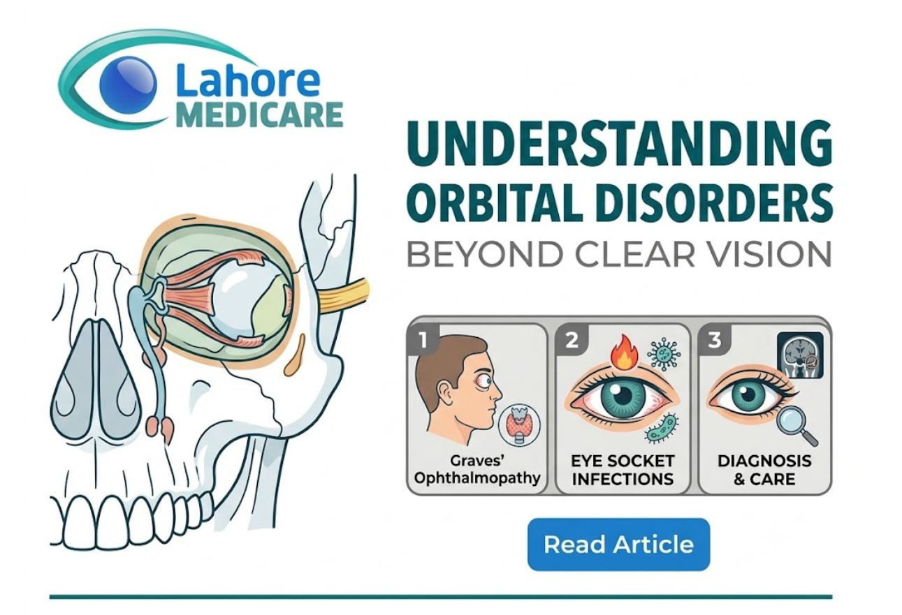
The orbital cavity (eye socket) is the bony cavity that encloses the bulb and accessory organs of the eye, including the ocular muscles, lacrimal glands, nerves, vessels, and retrobulbar adipose tissue. Every anatomical component housed within this bony socket fulfills a clear, dedicated role:
- Ocular Muscles: Six coordinated extraocular muscles that dictate smooth, multi-directional tracking movements.
- Lacrimal Glands: Glandular structures located in the upper outer orbit that produce tears to maintain smooth ocular lubrication.
- Nerves and Blood Vessels: Critical pathways, including the optic nerve, that transmit sensory data to the brain and supply oxygenated blood to surrounding tissue.
- Retrobulbar Adipose Tissue: Supportive fatty tissue surrounding the eyeball that acts as a natural shock-absorbing cushion against physical impacts.
Because these sensitive tissues share a fixed volume inside the skull, any disease process that causes tissue growth or fluid accumulation increases pressure on neighboring structures.
Primary Types of Orbital Cavity Disorders
Pathological changes within the eye socket stem from several clinical triggers, ranging from systemic autoimmune responses to localized bacterial invasions and primary cell growths.
Graves’ Ophthalmopathy (Thyroid Eye Disease)
Graves’ ophthalmopathy is an autoimmune disorder where the body’s immune system targets the soft tissues and extraocular muscles inside the eye socket. This targeted immune attack leads to chronic inflammation, muscle enlargement, and fatty tissue expansion. As the tissue volume expands within the non-yielding bony orbit, the eyeball is displaced forward, leading to significant ocular surface exposure and deep socket aching.
Orbital Cellulitis
Orbital cellulitis is an acute, severe bacterial infection affecting the deep fat and muscle tissues behind the eye. It frequently develops when a sinus infection spreads through the thin bony partitions separating the nasal sinuses from the orbit. Patients often experience sudden localized pain, fever, eyelid edema, and restricted eye tracking. Because the infection can rapidly compress the optic nerve, immediate hospital intervention is mandatory.
Rhabdomyosarcoma and Orbital Mass Lesions
Rhabdomyosarcoma is a fast-growing malignant tumor derived from immature skeletal muscle cells, occurring most frequently in pediatric patients. In addition to primary soft tissue sarcomas, benign vascular malformations, cysts, or secondary metastatic lesions can occupy physical space within the eye socket, displacing neighboring structures.
Lacrimal System Disturbances
Pathology involving the tear drainage system, such as acute inflammation or blockage of the lacrimal sac (dacryocystitis), creates painful swelling near the inner angle of the eye. These obstructions cause persistent tearing, localized tenderness, and recurring eye infections.
Recognizing Symptoms of Orbital Diseases
When disease processes disrupt the internal equilibrium of the eye socket, characteristic physical symptoms develop. The two most prominent clinical signs include:
- Exophthalmos (Proptosis): An abnormal protrusion or bulging of one or both eyes caused by swelling, fluid buildup, or expanding mass tissue behind the globe.
- Diplopia (Double Vision): Seeing double when tracking objects, caused by swollen, stiffened, or nerve-impaired extraocular muscles that can no longer realign both eyes smoothly.
Additional symptoms include constant deep orbit pressure, eyelid retraction, localized redness, and progressive vision loss if the optic nerve suffers physical compression. Seeking a comprehensive evaluation at a specialized center like Lahore Medicare provides access to the necessary imaging and clinical expertise required to protect your visual pathways.
Diagnostic Procedures and Treatment Protocols
Diagnosing conditions within the eye socket requires a multi-faceted approach. Ophthalmologists and orbital specialists rely on high-resolution computed tomography (CT) scans and magnetic resonance imaging (MRI) to evaluate soft tissue changes, measure muscle thickness, and map out bony structures.
Treatment modalities are tailored directly to the diagnosed condition:
- Medical and Conservative Management: Bacterial orbital cellulitis requires prompt intravenous antibiotic therapy in a clinical setting. Inflammatory conditions like thyroid eye disease are managed using systemic corticosteroids, targeted biological agents, or specialized orbital radiation to reduce tissue swelling.
- Surgical Interventions: When extreme tissue pressure threatens optic nerve health or causes severe eye protrusion, specialists perform orbital decompression surgery. This procedure removes thin sections of bony walls or excess retrobulbar fat to create extra space for swollen tissues.
- Oncological Therapies: Malignant conditions such as rhabdomyosarcoma are treated through coordinated protocols combining chemotherapy, targeted radiation therapy, and surgical resection when appropriate.
For individuals navigating inflammatory conditions or structural socket abnormalities, obtaining expert orbital disease treatment and management ensures that vision preservation remains the top medical priority. When surgical intervention or tissue biopsies are required, utilizing advanced ophthalmic diagnostic and surgical procedures helps restore proper socket mechanics and protects overall ocular health.
Frequently Asked Questions
What is the main difference between orbital cellulitis and preseptal cellulitis?
Preseptal cellulitis affects only the superficial skin and soft tissues of the eyelid, whereas orbital cellulitis involves the deeper tissues within the eye socket behind the orbital septum, posing a direct threat to vision.
Why does thyroid disease cause bulging eyes?
In thyroid eye disease (Graves’ ophthalmopathy), autoantibodies cause inflammation and swelling of the muscles and fat behind the eye. Because the bony socket cannot expand, the eye is pushed forward, causing it to bulge.
What causes diplopia in orbital disorders?
Diplopia, or double vision, occurs when inflammation, muscle swelling, or nerve compression prevents the extraocular muscles from moving both eyes together in perfect alignment, creating two separate visual images.
Is orbital cellulitis considered a medical emergency?
Yes. Orbital cellulitis is a sight-threatening and potentially life-threatening emergency. Immediate hospitalization and high-dose intravenous antibiotics are required to prevent permanent optic nerve damage or intracranial spread.
What is the role of retrobulbar fat in the eye socket?
Retrobulbar adipose tissue fills the space behind the eyeball, acting as a flexible protective cushion for the optic nerve, blood vessels, and extraocular muscles during routine eye movements.
How is orbital decompression surgery performed?
Orbital decompression involves removing small portions of the bone forming the socket walls, along with excess fatty tissue. This creates expanded space for swollen tissues, relieving pressure on the optic nerve and reducing eye protrusion.
What are the primary symptoms of rhabdomyosarcoma in the orbit?
Rhabdomyosarcoma typically presents as a rapidly progressing, painless or slightly tender bulging of the eye (proptosis) in children, often accompanied by eyelid swelling, redness, and restricted eye movement.
Can orbital diseases affect both eyes at the same time?
Yes. Autoimmune conditions like Graves’ ophthalmopathy frequently affect both eye sockets simultaneously, whereas bacterial infections, trauma, and localized tumors typically present on one side (unilaterally).
How do doctors view tissues inside the eye socket?
Specialists utilize detailed imaging tools like CT scans to examine the bony borders and MRI scans to evaluate soft tissues, ocular muscles, the optic nerve, and potential fluid or mass accumulations.
When should you consult a doctor for eye socket pain?
You should seek immediate medical evaluation if eye socket pain is accompanied by sudden double vision, visible eye protrusion, fever, eyelid redness, or a noticeable decline in visual acuity