The thought of losing your eyesight gradually without noticing any warning signs is a frightening concept, yet it represents the daily reality of a condition known as the “silent thief of sight.” Many individuals assume that an eye disease would announce itself through sharp pain, sudden blurriness, or obvious redness. However, certain chronic ocular conditions behave completely differently, masking their progression until irreversible harm has altered your field of vision.
The condition can be difficult to diagnose because symptoms don’t appear straight away – instead, they develop slowly over many years. This means many patients only seek treatment when they notice they’re losing their sight, when significant damage has already occurred. Understanding how this asymptomatic progression occurs is the first step toward protecting your long-term optical health and ensuring your eyes remain functional for life.
Why Internal Eye Fluid Causes Silent Nerve Damage
To understand how this condition progresses without a single early warning sign, it helps to look at the continuous biological processes occurring inside the eye. Your eyes naturally produce a clear fluid to shape, nourish, and cleanse internal structures. In a perfectly healthy eye, an equal amount of this fluid drains out through microscopic channels at the same rate it is produced, keeping internal pressure stable.
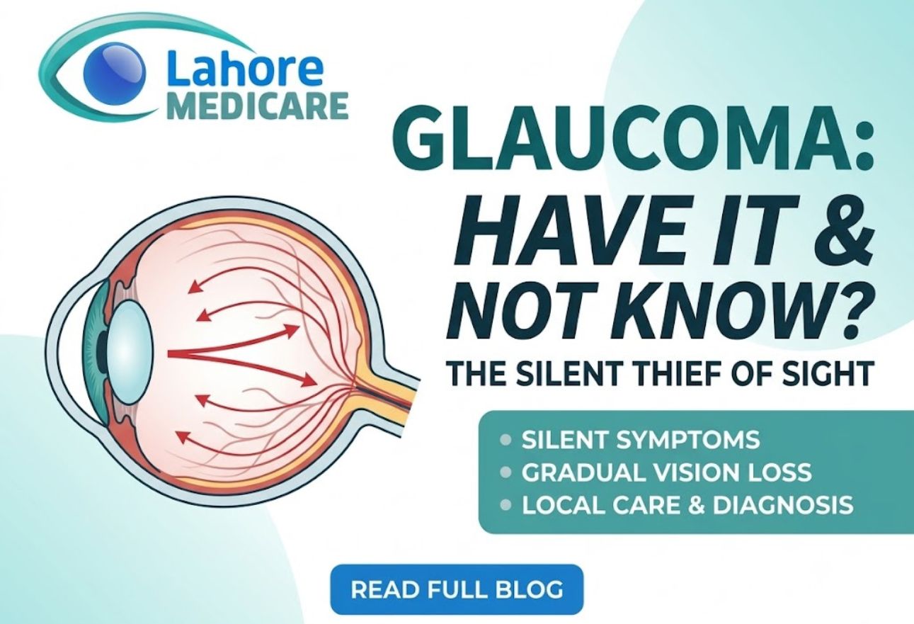
Glaucoma is a group of conditions caused by normal fluid in the eye that hasn’t drained properly. This creates pressure that damages the optic nerve connecting the eye to the brain, resulting in sight loss. Because this pressure build-up happens gradually, the delicate nerve fibers at the back of the eye are compressed and destroyed one by one. The brain easily adapts to the initial micro-losses in your peripheral vision, filling in the blind spots so effectively that you feel completely normal while the structural breakdown continues. For residents looking to catch these changes before they disrupt your life, scheduling a regular screening for Glaucoma Treatment in lahore remains the most effective defense against permanent vision impairment.
Assessing Your Personal Risk Factors
Medical researchers continue to study the precise molecular triggers behind drainage failures, but the core systemic vulnerabilities are well-documented. Although it’s not clear exactly why this happens, factors such as age, family history, racial background and other medical conditions such as diabetes and short-sightedness can increase the risk. It can affect people of all ages, but it’s most common in adults.
| Risk Factor | Impact on Eye Health |
| Family History | Increases genetic predisposition to drainage channel narrowness. |
| Medical Conditions | Chronic illnesses like diabetes alter blood flow and weaken ocular vessels. |
| Refractive Errors | High short-sightedness physically elongates the eye, straining the optic nerve. |
Because these risk factors combine silently, individuals over forty or those living with chronic metabolic conditions must treat eye exams as a fundamental part of their healthcare routine. Relying purely on how well you can see a digital screen or road sign today will not reveal whether internal fluid dynamics are slowly compromising your nerve pathways.
Managing a Progressive Eye Condition for Life
When a detailed ophthalmic evaluation reveals structural changes or elevated internal pressure, the primary goal shifts from diagnosis to preservation. There are different types of glaucoma, and treatment will depend on the type a patient has. Glaucoma can’t be cured, and vision that has already been lost cannot be restored. But further sight loss can be prevented via medication or surgery. Each glaucoma patient requires lifelong management for best results.
Modern care centers focus heavily on slowing down or stopping the progression of nerve compression. Specialized prescription eye drops work by either reducing the amount of fluid your eye generates or opening up natural exit pathways to increase outflow. When daily medications are not sufficient to keep your internal pressure within a safe, stable range, advanced medical interventions become necessary. Exploring the appropriate clinical Procedures of treatment For Glaucoma patients allows specialists to create alternative drainage routes using precise laser treatments or delicate micro-surgical techniques.
Breaking the Barrier to Early Local Diagnosis
The greatest obstacle in modern ophthalmology is not a lack of effective therapeutic tools, but rather the delayed timing of patient arrivals. Yet the challenge is that patients often don’t seek treatment for glaucoma until it is too late to save their sight. This is why we are working with partners in Africa to introduce pilot programmes to prevent and treat glaucoma. These international public health efforts focus on taking advanced diagnostic equipment directly into underserved communities where routine clinical visits are rare.
Closer to home, the focus centers on establishing accessible, high-quality diagnostic infrastructure within urban hubs and regional clinics. Our aim is to make sure glaucoma is diagnosed and treated as part of local eye health services, to ensure patients’ sight can be saved. By integrating specialized tonometry, the test used to measure internal eye pressure into routine community wellness packages at centers like Lahore Medicare, local families can identify pressure spikes decades before peripheral vision failure begins, changing the trajectory of their visual health forever.
Frequently Asked Questions
Can I look in a mirror and see signs of eye pressure?
No. Chronic elevated eye pressure does not cause eye redness, pupil changes, or visible swelling on the outside. It can only be detected by a specialist using specialized clinical diagnostic tools.
Why is optic nerve damage from eye fluid permanent?
The optic nerve is made of delicate central nervous system fibers. Once these specific nerve cells are crushed by prolonged fluid pressure, they cannot regenerate or repair themselves, making early prevention vital.
Does high blood pressure cause high eye pressure?
While they are separate medical issues, systemic high blood pressure can influence fluid production and blood flow dynamics inside your eye, indirectly elevating your long-term risk for nerve damage.
How often should a diabetic patient check for glaucoma?
Diabetic individuals should undergo a comprehensive, dilated eye exam at least once every year. Diabetes affects ocular blood vessels, making the optic nerve more vulnerable to fluid pressure changes.
What happens during a standard eye pressure test?
A specialist uses a quick, painless device called a tonometer to gently measure the resistance of your cornea. This reading tells the consultant exactly how much fluid pressure is pushing outward.
Are children completely safe from developing glaucoma?
No. While it is most common in aging adults, congenital or juvenile forms can affect infants and children due to improper development of the eye’s natural drainage pathways from birth.
Can prescription eye drops restore my lost peripheral vision?
No. Eye drops are strictly designed to lower internal fluid pressure to protect your remaining healthy nerve fibers. They cannot reverse or cure the blind spots that have already formed.
What is the difference between open-angle and acute glaucoma?
Open-angle develops slowly and painlessly over many years without immediate symptoms. Acute angle-closure is a sudden, painful medical emergency caused by a total drainage blockage requiring immediate care.
Can regular exercise help lower internal eye fluid pressure?
Regular, moderate aerobic exercise like walking or jogging has been shown to temporarily lower eye pressure in some individuals, but it must never replace your prescribed medical treatments.
Why does short-sightedness increase my structural risk?
Severe short-sightedness physically stretches the back layers of the eye. This elongation structural weakens the tissue around the optic nerve, making it more sensitive to normal fluid pressure.