
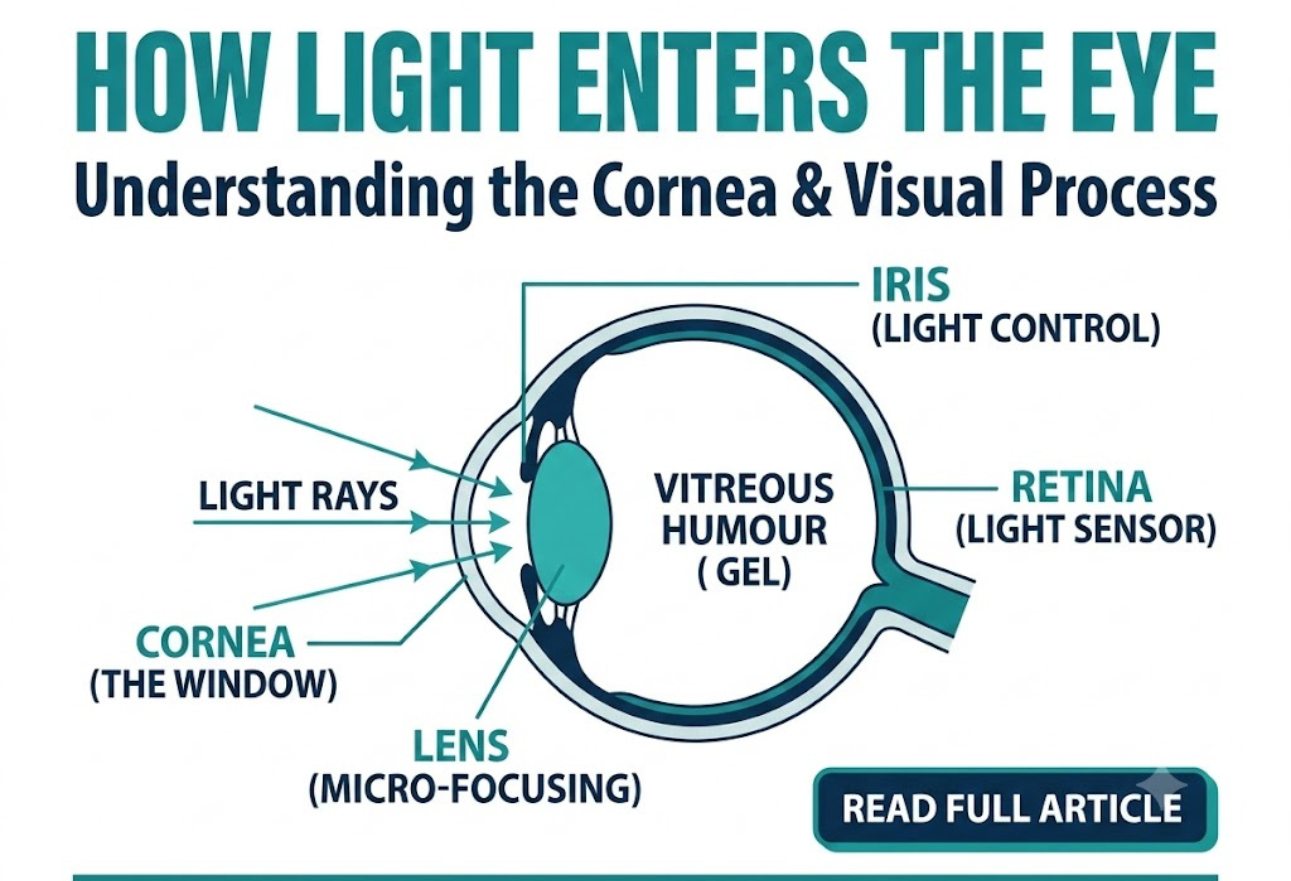
Every sight we experience, from the vibrant colors of a sunset to the sharp text on a smartphone screen, relies entirely on a beautiful biological system. The images we see are made up of light reflected from the objects we look at. To convert this wandering light into a crisp mental picture, the human eye coordinates an intricate pathway of clear lenses, protective layers, and light-sensitive cells. Understanding this journey reveals exactly how our eyes build our visual reality and why protecting each stage of the process is so critical.
The Entry Point: The Cornea and Light Regulation
The visual process begins the exact millisecond light bounces off an object and moves toward your face. This light enters the eye through the cornea, which acts like a window at the front of the eye. Because this outermost layer is directly exposed to the external world, keeping it smooth and transparent is paramount for clear vision.
Once inside, the amount of light entering the eye is controlled by the pupil, which is surrounded by the iris – the coloured part of the eye. The iris functions dynamically like the aperture of a professional camera lens, expanding in dim environments to gather more light and constricting under bright glare to prevent internal damage. Because the front part of the eye is curved, it bends the light, creating an upside down image on the retina. It is then up to the neurological system to finalize this information, as the brain eventually turns the image the right way up.
When an underlying medical issue or physical scratch distorts this clear front surface, the light passing through becomes irregular, resulting in distorted or highly blurred vision. For individuals struggling with structural irregularities or progressive scarring on this protective outer layer, seeking specialized Corneal Disease Treatment in lahore is an essential step to restoring the eye’s natural entry window.
The Micro-Focusing System: The Lens and Internal Humours
After passing through the pupil, the light rays must travel deeper into the eye through a series of fluids and adjustable structures that sharpen the image focus. Immediately behind the iris sits a clear liquid called the aqueous humour, which fills a small chamber behind the cornea to provide essential nutrients and maintain proper structural pressure.
Directly following this chamber is the lens, a clear disc-like structure that helps to focus light on the retina. The lens is uniquely dynamic compared to the rigid cornea. It can do this because it is adjustable, and uses a muscle called the ciliary muscle to change shape and help us focus on objects at different distances. When looking at a close object, the ciliary muscle flexes, allowing the lens to thicken and bend light more sharply; when viewing the horizon, it relaxes. This automatic focusing of the lens is a reflex response and is not controlled by the brain, operating instantly without conscious thought.
Once through the lens, the light travels through the vitreous humour, the clear gel that fills the space between the lens and the retina. This gel maintains the spherical shape of the eye, ensuring that the light rays hit the back wall precisely where they should.
The Retina: Converting Light Into Brain Signals
The ultimate destination for these focused light rays is the retina, a complex part of the eye, and its job is to turn light into signals about images that the brain can understand. Only the very back of it is light sensitive: this part of the retina is roughly the area of a 10p coin, and is packed with photosensitive cells called rods and cones.
These millions of specialized photoreceptors are divided into two distinct teams, each designed for entirely different lighting environments:
- Cones: These are the cells responsible for daylight vision. There are three kinds, each responding to a different wavelength of light: red, green and blue. The cones enable us to see images in colour and detail, operating exclusively in moderate to bright light conditions.
- Rods: These are responsible for night vision. They are sensitive to light but not to colour, meaning that in darkness, the cones do not function at all, leaving the rods to map out our surroundings in grayscale shades.
Once the image is clearly focused on the sensitive part of the retina, energy in the light that makes up that image creates an electrical signal. Nerve impulses can then carry information about that image to the brain through the optic nerve, where it is instantly interpreted into the full-color world we experience.
Supporting Structures and Vision Longevity
The intricate process of focusing light cannot occur without a robust internal infrastructure supporting these sensitive tissues. Protecting the rear layers of the eye are the sclera and the choroid. The sclera is the white part of the eye, forming an outer layer that protects everything inside from external trauma. Meanwhile, the choroid is the layer of the eye that lies between the retina and the sclera. It is made up of layers of blood vessels that nourish the back of the eye, supplying the oxygen and nutrients needed to keep the rods and cones alive.
Maintaining the balance of all these complex components requires regular specialized checkups. At Lahore Medicare, comprehensive diagnostic exams evaluate everything from the clarity of your cornea to the nerve integrity of your retina. If structural damage or progressive blurring does begin to compromise your sight, exploring modern clinical Procedures of treatment with an eye care professional ensures that each delicate step of the visual pathway continues to function perfectly for years to come.
Frequently Asked Questions
Why does the eye create an upside-down image on the retina?
Because the surface of the cornea and the internal lens are naturally curved, they act like a convex lens. This curve bends the incoming light rays across each other, which naturally flips the projected image upside down by the time it reaches the retina.
What is the exact role of the cornea in vision?
The cornea acts like a protective front window for the eye. Its curved structure provides the primary focusing power, bending incoming light rays so they can pass cleanly through the pupil and converge accurately onto the lens.
How do rods and cones differ in dark rooms?
Cones require bright light to detect colors and fine details, so they do not function at all in darkness. Rods are incredibly sensitive to low light levels and handle your night vision, though they cannot perceive different colors.
What changes the shape of the internal eye lens?
The lens relies on the ciliary muscle to adjust its shape. When focusing on close objects, this muscle contracts to make the lens rounder; when focusing at a distance, it relaxes to flatten the lens. This happens completely as an automatic reflex.
What is the function of the choroid layer?
The choroid is a vital layer packed with blood vessels located between the retina and the white sclera. Its primary responsibility is to provide constant nourishment, oxygen, and blood flow to the sensitive outer layers of the retina.
How does the iris control the amount of light entering?
The iris is the colored muscle structure that surrounds the pupil. In bright light, the muscles contract to shrink the pupil opening, while in dark environments, they relax to widen the pupil, regulating light entry.
What is the difference between aqueous and vitreous humour?
Aqueous humour is a thin, watery liquid found in the small chamber directly behind the cornea. Vitreous humour is a thicker, clear gel-like substance that fills the large main space between the lens and the back retina.
Why is the optic nerve critical for our sight?
The optic nerve acts as the primary data cable for vision. Once the retina converts focused light into electrical nerve impulses, the optic nerve carries these raw signals directly to the brain to be translated into conscious images.
Can a damaged cornea fix itself over time?
Minor surface scratches on the cornea can often heal quickly because the outer layer grows back rapidly. However, deep injuries, infections, or diseases can cause permanent scars that require specialized medical treatments to clear.
What part of the retina is most sensitive to light?
The most light-sensitive area is located at the very back of the retina, spanning an area roughly the size of a 10p coin. This specialized region is highly dense with photoreceptors to capture maximum image detail and color.