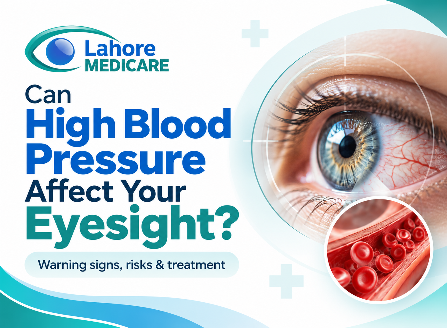
High blood pressure (hypertension) is often called a “silent killer” because it damages blood vessels throughout the body without causing obvious initial symptoms. While many people associate hypertension primarily with heart disease and stroke, its impact on visual health is equally profound. The retina, the light-sensitive layer of tissue at the back of the eyeball, relies on a dense, delicate network of microvascular blood vessels to function. When systemic blood pressure remains consistently elevated, these tiny vessels undergo structural damage that can lead to permanent vision impairment or total sight loss.
Understanding how systemic hypertension affects the ocular microvasculature, recognizing early warning indicators, and seeking regular ophthalmic screenings are essential steps in protecting long-term vision.
How Hypertension Damages Ocular Blood Vessels
The delicate microvascular network supplying the retina, optic nerve, and choroid is particularly susceptible to systemic pressure shifts. Uncontrolled high blood pressure leads to distinct physical changes within the intraocular circulation:
- Arteriolar Narrowing and Sclerosis: Persistent pressure causes the muscular walls of retinal arterioles to thicken and stiffen. Under an ophthalmoscope, these thickened vessels reflect light abnormally, producing classic clinical signs known as “copper wiring” or “silver wiring.”
- Vascular Leakage and Hemorrhage: As arteriolar walls weaken, fluid, lipids, and blood leak into surrounding retinal tissue. This results in retinal edema, flame-shaped hemorrhages, and hard lipid deposits.
- Ischemia and Cotton-Wool Spots: When damaged microvessels become completely blocked, localized patches of the retina are deprived of oxygen. These ischemic areas swell and manifest clinically as soft, fluffy white patches called “cotton-wool spots.”
Ocular Conditions Caused by High Blood Pressure
Hypertension can trigger several severe sight-threatening ocular conditions:
1. Hypertensive Retinopathy
Hypertensive retinopathy is progressive damage to the retinal microvasculature caused by elevated blood pressure. It is graded from mild arteriolar narrowing (Grade 1) to severe optic nerve swelling or papilledema (Grade 4). In advanced stages, retinal swelling and widespread hemorrhages cause significant blurred vision and visual field loss.
2. Retinal Artery and Vein Occlusions
Elevated blood pressure increases the risk of blood clots and vascular blockages inside the eye, commonly known as an “eye stroke”:
- Retinal Vein Occlusion (RVO): Stiffness in adjacent arterioles compresses the retinal vein, causing blood and fluid to back up, leading to sudden, painless visual loss.
- Retinal Artery Occlusion (RAO): A blockage in the main retinal artery deprives the retina of oxygenated blood, resulting in immediate and severe sight loss.
3. Hypertensive Choroidopathy and Optic Neuropathy
In severe or malignant hypertension, fluid leakage can extend beneath the retina into the vascular choroid layer (choroidopathy), leading to retinal detachment. Additionally, restricted blood flow to the optic nerve head (ischemic optic neuropathy) causes permanent nerve cell death and central visual field loss.
Evaluating retinal vascular health alongside complex cardiovascular eye diseases and conditions ensures early clinical identification before irreversible tissue damage occurs.
Recognizing Symptoms and Warning Signs
In its early stages, hypertensive retinopathy produces virtually no noticeable visual symptoms. As microvascular damage advances, patients may experience:
- Sudden or Gradual Blurry Vision: Reduced visual sharpness caused by fluid accumulation (edema) in the central macula.
- Double Vision (Diplopia): Occurs when severe hypertension impairs cranial nerves controlling the extraocular muscles.
- Dim or Partial Vision Loss: Shadowy or dark patches in the visual field caused by retinal vein blockages or localized ischemia.
- Sudden Total Sight Loss: Severe visual deficit resulting from a major retinal artery occlusion or optic nerve infarction.
Because microvascular changes often develop silently, scheduling comprehensive dilated eye exams at an established center like Lahore Medicare allows specialists to detect early vascular damage before symptoms manifest.
Treatment and Medical Management
Managing ocular complications from high blood pressure requires a collaborative approach involving primary care physicians, cardiologists, and ophthalmologists:
- Systemic Pressure Control: The primary defense against advancing ocular damage is lowering systemic blood pressure through lifestyle modifications, dietary adjustments, and antihypertensive medications.
- Anti-VEGF Injections: Intravitreal injections of Vascular Endothelial Growth Factor inhibitors help reduce macular swelling and control abnormal fluid leakage in secondary retinal vein occlusions.
- Retinal Laser Photocoagulation: Targeted laser therapy seals leaking microvessels and shrinks ischemic tissue to prevent recurrent intraocular bleeding.
Patients requiring advanced diagnostic imaging, laser photocoagulation, or intravitreal therapies can access advanced ophthalmic procedures to protect fragile retinal circulation and maintain functional vision.
Frequently Asked Questions
Can high blood pressure cause sudden permanent blindness?
Yes. Severe or uncontrolled high blood pressure can cause a sudden, painless loss of vision through a retinal artery occlusion, a central retinal vein occlusion, or an ischemic optic nerve stroke. In these emergency situations, the blockage deprives the retina or optic nerve of vital oxygenated blood, which can lead to permanent structural tissue death and permanent blindness if not evaluated immediately by an ophthalmologist.
What are the early warning signs of hypertensive retinopathy?
In its earliest phases, hypertensive retinopathy usually causes no obvious visual symptoms at all, which is why it is often called a silent condition. As microvascular damage progresses, early warning signs can include subtle bouts of blurry vision, mild visual field distortions, dim vision, double vision, or frequent headaches. A dilated eye exam is the most reliable way to catch these vascular changes before permanent vision loss occurs.
Can vision loss caused by high blood pressure be reversed?
Reversibility depends heavily on the extent and duration of the damage. Early-stage hypertensive retinopathy changes, such as mild retinal swelling or small hemorrhages, can often resolve and vision can improve once systemic blood pressure is strictly controlled. However, if long-standing hypertension has caused optic nerve atrophy, permanent macular scarring, or tissue death from an eye stroke, the resulting vision loss is usually permanent.
How does an ophthalmologist detect high blood pressure during an eye exam?
During a routine dilated fundus examination, an eye specialist uses a specialized slit-lamp microscope and magnifying lenses to directly view the living retinal blood vessels. They check for telltale structural signs of vascular damage, such as narrowed or stiffened arterioles, light reflection changes known as copper or silver wiring, localized cotton-wool spots, micro-hemorrhages, and swelling of the optic nerve head.
How often should someone with high blood pressure get their eyes checked?
Individuals diagnosed with systemic hypertension should undergo a comprehensive dilated eye examination at least once a year. If you have already been diagnosed with hypertensive retinopathy, diabetic eye complications, or a history of retinal vein occlusions, your eye care specialist may recommend more frequent monitoring screenings every three to six months to closely track vascular health and evaluate treatment progress.
What is the difference between an eye stroke and a brain stroke?
An eye stroke occurs when elevated blood pressure, a blood clot, or plaque blocks the blood flow specifically within the retinal arteries, retinal veins, or optic nerve, causing sudden localized vision loss. A brain stroke occurs when a blood vessel supplying brain tissue becomes blocked or ruptures, leading to widespread neurological symptoms such as facial drooping, arm weakness, speech difficulty, and potential systemic paralysis.
Can lowering my blood pressure improve my eye damage?
Yes, bringing your systemic blood pressure into a normal target range through lifestyle changes and prescribed antihypertensive medications can significantly halt the progression of retinal vascular damage. Over time, controlling your blood pressure allows leaking retinal fluid to reabsorb, reduces retinal swelling, and lowers the future risk of developing severe complications like retinal vein occlusions or hypertensive choroidopathy.
What is copper wiring and silver wiring in hypertensive retinopathy?
Copper wiring and silver wiring are clinical terms used by eye specialists to describe the altered visual appearance of retinal arterioles under high blood pressure. As chronic hypertension thickens and stiffens the muscular walls of retinal blood vessels, the central light reflex changes from a normal thin red line to a broader, shiny copper sheen. In advanced stages, the vessel wall becomes so opaque that it resembles a reflective silver wire.